In many rural regions, family poultry farming is key to food security and the economy, as it is accessible to the whole family and compatible with other agricultural activities. However, it is at risk due to the loss of local chicken breeds suitable for raising in rustic conditions.
- In addition, ornamental and fighting bird breeding faces health problems due to the lack of adequate technologies, which creates challenges for field veterinarians who must diagnose serious diseases.
- Two neoplastic viral diseases, Marek’s disease and Lymphoid Leukosis, represent major threats.
Marek’s disease causes tumor infiltrations in various organs and presents variable nervous symptoms, while lymphoid leukosis mainly affects the bursa of Fabricius, liver, spleen, kidney, and ovary, but does not infiltrate the peripheral nerves. It is essential to train rural veterinarians to protect non-technified poultry farming.
ETIOLOGY OF MAREK’S DISEASE
- The virus that causes Marek’s disease is an alpha herpesvirus belonging to the genus Mardivirus.
- The viral particles are enveloped and measure 150-160 nm in diameter.
- The genome consists of a double strand of DNA, and the nucleocapsid is hexagonal with 162 capsomeres.
Previously, alphaherpesviruses were classified into three serotypes, but currently three species are recognized:
- Gallid alphaherpesvirus 2 which is the etiological agent of Marek’s disease (formerly serotype 1), with low and high virulence and attenuated strains.
- Gallid alphaherpesvirus 3 (formerly serotype 2). It infects chickens but is avirulent and therefore non-oncogenic.
- Meleagrid alphaherpesvirus 1, (Herpesvirus in turkeys, formerly serotype 3). Infects turkeys but is avirulent and non-oncogenic.
Gallid alphaherpesvirus 2 has four pathotypes:
- Medium virulence (mEM).
- Virulent (vEM).
- Very virulent (vvEM).
- Very virulent plus (vv+EM).


ETIOLOGY OF LYMPHOID LEUKOSIS
The Lymphoid Leukosis is caused by a Type C oncovirus belonging to the Retroviridae. This virus has:
- Protein envelope encoded by the gag gene, which contains the group-specific antigen (gsa), useful for diagnosis.
- Second outer envelope encoded by the env gene.
- Genome composed of two RNA segments (35S) and a reverse transcriptase inside, encoded by the pol gene.
Avian leukosis viruses are grouped into subgroups A, B, C, D, and E:
- A, B, C, and D: these are exogenous, transmitted horizontally or congenitally.
- E: is endogenous, is transmitted genetically as integrated provirus into the host’s DNA.
In 1991, Virus J (ALV J) was identified, which affects heavy-type birds and causes early neoplasms, specifically myelocytomatosis.
- Subgroups A and B are the most common and the main causes of lymphoid leukosis.
TRANSMISSION OF MAREK’S DISEASE AND LYMPHOID LEUKOSIS
- Marek’s disease: transmitted horizontally through dust containing flakes from the feather follicles, where the infectious virions are found.
- Lymphoid Leukosis: can be transmitted horizontally or vertically:
- Congenital vertical: the embryo becomes infected in the hen’s oviduct. This route is mainly caused by subgroups A and B.
- Vertical genetic: the provirus is transmitted through gametes, both from the rooster and the hen. Involves endogenous subgroups E and J.
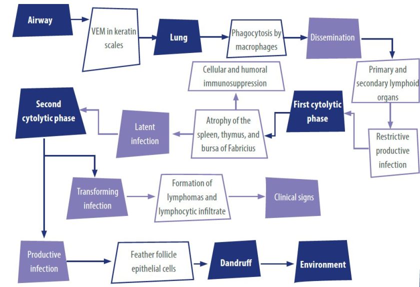
PATHOGENESIS OF MAREK’S DISEASE
Marek’s disease begins when birds inhale viral particles present in dust contaminated with flakes from feather follicles.
- These particles reach the lungs, where they are captured by macrophages that transport them to the lymphoid organs (spleen, thymus, and bursa of Fabricius).
- In these organs, the virus causes an initial cytolytic phase, causing atrophy of the lymphoid tissues and establishing a latent infection.
- Then, it can be presented a second cytolytic phase, which gives rise to two distinct processes:
- Neoplastic transformation of T lymphocytes, which generates lymphoid tumors.
- Infection of feather follicle epithelial cells, which produces complete viral particles without cell transformation but with the ability to transmit to other birds (Table 1).

PATHOGENESIS OF LYMPHOID LEUKOSIS
The avian leukosis virus does not have oncogenes, but its RNA is converted into Proviral DNA by reverse transcriptase, inserting itself into the genome of the infected cell.
- In chickens, virions have been isolated in the ovary and oviduct, especially in the magnum, where they infect the zygote during egg passage, causing congenital vertical transmission.
At birth, some embryos already have virions in the liver and kidney.
- Between the 6th and 8th week of life, giant lymphoid follicles can be detected in the Fabricius sac, indicating the onset of neoplastic transformation of B lymphocytes.
- At 10 weeks, macroscopic tumors appear in the sac, and metastases to other organs occur from the 20th week.
CLINICAL SIGNS OF MAREK’S DISEASE
The clinical signs of Marek’s disease are mainly nervous, characterized by progressive paresis, paralysis of one or more limbs, and incoordination, as well as anorexia, depression, paleness of combs and wattles, dehydration, and diarrhea leading to cachexia.
CLINICAL SIGNS OF LYMPHOID LEUKOSIS
In lymphoid leukosis, the main clinical signs are anorexia, cachexia, depression, ruffled feathers, paleness or cyanosis of combs and wattles, and a distended abdomen.
- An increase in the tissue mass of the Fabricius sac and liver can sometimes be detected by palpation.


LESIONS IN MAREK’S DISEASE
The lesions of Marek’s disease consist of five types of tumor cell infiltrations, which are:
- Visceral: Ivory-white or grayish nodules in organs such as the liver, kidney, spleen, heart, ovary, proventriculus, and lungs. They can be observed from the fourth week of age in acute cases, without involving the spinal cord or peripheral nerves.
- Cutaneous: Protruding masses in the feather follicles.
- Muscular: White nodular masses in the pectoral muscles, although these are rare.
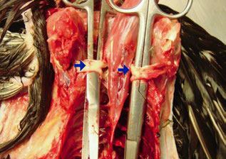
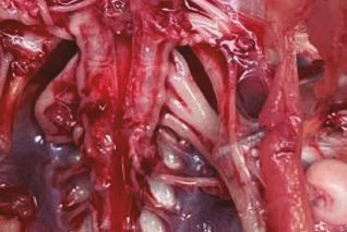
- Neural: Affects between 20-40% of birds and is the most common form. The lesions are unilateral and are found in nerve plexuses ( brachial, lumbosacral, sciatic). The affected nerves thicken, turning yellow or pearly gray and losing their striations. The mesenteric and cranial nerves may also be affected.
- Ocular: These are not described in detail, but usually include changes in the iris and other eye tissues, which are typical of the disease.
LYMPHOID LEUKOSIS LESIONS
The characteristic lesions of lymphoid leukosis are mainly tumors that occur in various viscera. The most important points are:
Characteristics of tumors:
They are nodular in shape and are white, yellow, or gray.
Location of tumors:
The tumors occur most frequently in the following organs:
- Liver
- Spleen
- Heart
- Kidneys
- Lungs
- Ovary
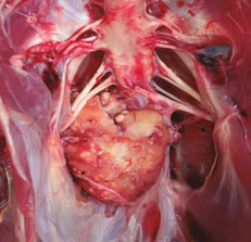
- Fabricius sac (almost always affected).
Tumors in the Fabricius sac:
- The early tumors can be detected from 10 weeks of age.
- They are characterized by a marked thickening of one or more folds of the inner wall of the sac.
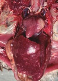
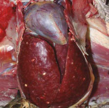
Liver lesions:
- In the liver, tumor lesions may be infiltrative rather than nodular.
- They manifest as marked hepatomegaly (enlargement of the liver). (Figures 4 and 5)


In this first part, we presented the general aspects of Marek’s disease and lymphoid leukosis. In the second part, we will address the histopathology, diagnosis, prevention, and control measures for these diseases in family poultry farming.