In the first part of this article, the importance of family poultry farming in rural areas and the health challenges it faces, especially due to viral diseases such as Marek’s Disease and Lymphoid Leukosis, was addressed.
- Both present similar clinical manifestations and anatomopathological lesions, making differential diagnosis difficult and affecting the health of the flocks.
This second part delves into the histopathological, diagnostic, and prevention and control strategies.
The objective is to provide rural veterinarians with technical tools to protect native birds and ensure the sustainability of family poultry farming.
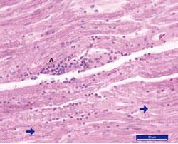
HISTOPATHOLOGY OF MAREK’S DISEASE
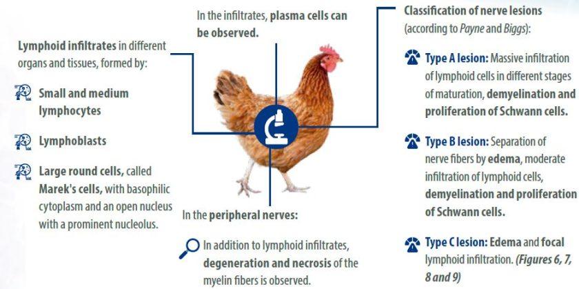
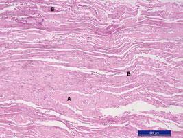
The histological lesions in Marek’s disease are characterized by:


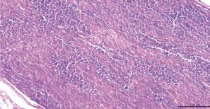

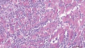


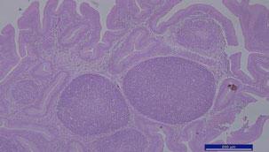
HISTOPATHOLOGY OF LYMPHOID LEUKOSIS
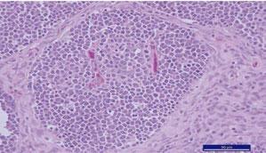
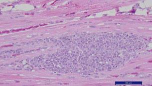
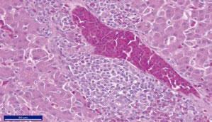
The microscopic lesions in lymphoid leukosis consist of homogeneous infiltrates of lymphoblastic cells.
- The neoplasms originate in the bursa of Fabricius, in the follicles, where there is a tumoral transformation of B lymphocytes.
- This occurs around the sixth week of age. These are large, very prominent tumoral follicles that compress and displace the normal follicles. (Figures 10, 11, 12 and 13).


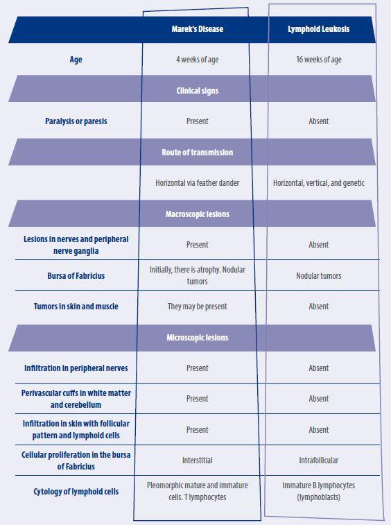
COMPARATIVE TABLE MAREK VS LEUKOSIS

DIAGNOSIS IN MAREK’S DISEASE
Clinical history:
- It is crucial to know whether the birds have been vaccinated against Marek’s Disease, which vaccine was used, and the vaccination schedule.
Complete necropsy:
- It must include the inspection of the nerve plexuses and peripheral nerves.
Histopathology:
- Histopathology is essential for an accurate diagnosis, especially depending on the type of lymphoid infiltration and its location.
- The lymphocytic infiltrates in the nerves are pathognomonic of the disease.
Diagnostic tests:
- Immunohistochemistry and PCR are useful for identifying the disease.
- Molecular techniques, such as quantitative PCR, are used to detect the viral genome in tumors.
Serological tests:
- The serological tests include:
- Immunofluorescence
- Immunohistochemistry
- ELISA
- Agar precipitin test
- Virus neutralization.
Previously, the key methods for diagnosing Marek’s Disease, from clinical history to specialized tests, are highlighted.
DIAGNOSIS OF LYMPHOID LEUKOSIS
Detection age:
- Lymphoid leukosis is not observable in broiler chicken flocks, since the detection of tumors is generally performed from 16 weeks of age onwards.
Differentiation from Marek’s Disease:
- It is important to emphasize the absence of lesions in the nerve plexuses and peripheral nerves, as these are pathognomonic of Marek’s Disease.
Histological characteristics:
- In lymphoid leukosis, the tumor cell infiltrates are uniform and are composed of lymphoblasts.
- In contrast, in Marek’s Disease, the lymphoid infiltrates are pleomorphic (they present different forms).
Tests for diagnosis:
- Several biological tests have been described for the detection of lymphoid leukosis, including:
- Virological and serological criteria to detect the virus, viral antigens, and specific antibodies.
- These tests are useful to ensure the absence of infection in pathogen-free birds and breeders. They also help to ensure that the vaccines are free of viruses.
Limitations of the tests:
- The diagnostic value of these tests is limited, since oncogenic viruses in birds frequently produce infection without inducing neoplasms.
PREVENTION AND CONTROL OF MAREK’S DISEASE
The biosecurity measures currently used in poultry farming influence the prevention of all infectious diseases, including Marek’s Disease; however, vaccination is essential and must be carried out on the first day of life or at 18 days in ovo.
Vaccines with the following serotypes are commercially available:
- Serotype 3 HVT: Strain FC-126.
- Serotype 2: Strains SB-1 and 301B/1
- Serotype 1: Attenuated strain CVI-988 and HPRS-16.
There are also bivalent vaccines such as:
- SB-1 (serotype 2) + FC-126 (serotype 3).
- CVI-988 (serotype 1) + HVT.
And trivalent vaccines such as:
- Serotype 1 + serotype 2 + serotype 3.
Among the recombinant vaccines are:
- HVT + IBD viral gene + ILT viral gene.
- HVT + IBD viral gene + VND viral gene.
PREVENTION AND CONTROL OF LYMPHOID LEUKOSIS
Likewise, implement all biosecurity measures and, if possible, permanently monitor breeder flocks with biological tests for viral detection, viral antigens, and specific antibodies, in order to eliminate birds carrying the virus or those presenting antibodies. There is no vaccination against avian leukosis.
CONCLUSION
- Marek’s Disease and Lymphoid Leukosis are serious threats to family poultry farming. Their control requires accurate diagnosis, strict biosecurity, and vaccination (only for Marek’s Disease).
- It is essential to train rural veterinarians and protect native birds to maintain avian health, preserve genetic diversity, and ensure the sustainability of production in non-technified systems.