Content available at: Español (Spanish)
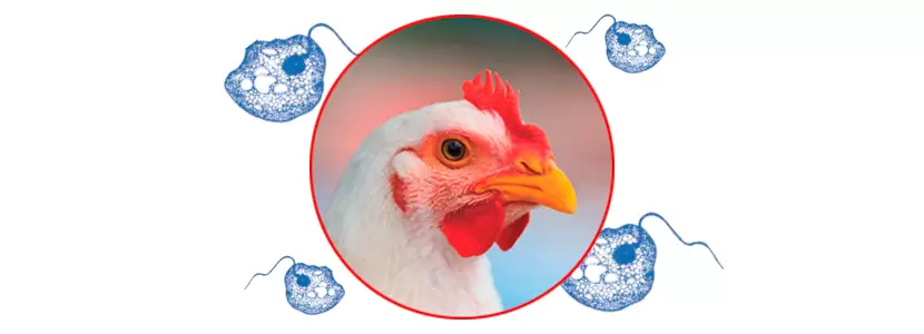
Histomoniasis, also known as blackhead disease is a complex entity. Although primarily affecting turkeys with lesions in the cecum and liver, it can also occur in laying and broiler breeder hens (where the lesions are often limited to the cecum only).
Histomoniasis is caused by the protozoan flagellate, Histomonas meleagridis (H. meleagridis), which has a broad range of hosts, infecting gallinaceous birds, including pheasants, partridges, and quails, as well as chickens and turkeys.
With the ban of most drugs used to fight the disease, and changes in animal husbandry like reusing litter and/or increased stocking density, histomoniasis has re-emerged in many areas, including North America and Europe. The approach to the control of histomoniasis is now based on prevention and the use of new diagnostic methods to better understand how to manage and eradicate the disease.
VECTORS & TRANSMISSION
Ingestion of the adult common cecal worm or nematode ( Heterakis gallinarum ) or its embryonated eggs infected with H. meleagridis .
Heterakis gallinarum is the only nematode known to serve as an intermediate host (or biological carrier) for histomoniasis . After a series of divisions, a very small and uniquely adapted form of H. meleagridis actively invades the reproductive tract of the caecal nematode and subsequently sheds off within the infected nematode egg.
Cecal nematode eggs are extremely resistant to the environment and can remain infectious in the litter or dirt floor for 2-3 years.
Egg transport of the cecal nematode infected by other mechanical carriers.
These include earthworms (which can ingest the eggs) or mechanical carriers of the disease like flies or rodents that can simply carry the sticky eggs on their bodies. PCR testing has shown that black beetles ( Alphitobius diaperinus ) may contain H. meleagridis DNA, providing evidence for its role as a possible carrier.
People and equipment (i.e. scales, transport cages, partitions, nets, vaccination tables, etc.) can also act as carriers of disease. Flood water during very humid times can act as a trigger for increased earthworm activity in particular areas.
Direct transmission of H. meleagridis through ingestion of liquid feces through the cloacal route known as cloacal consumption.
Tested only in turkeys, the cloacal route may be the reason why disease spread appears to be faster in turkeys compared to chickens.
Experimentally, direct oral inoculation of H. meleagridis has low success due to the susceptibility of the parasites to crop and gizzard acidity. However, histomoniasis manifests when H. meleagridis is administered to turkeys intra-cloacally or by “cloacal consumption”.
Outside the host (or the intermediate host: Heterakis gallinarum ), H. meleagridis has a low persistence and survival time , although it has been shown that it can survive up to 9 hours in water or fecal material. There is also some evidence that a cystic form can develop that would allow protozoa to survive for extended periods in the environment, although this has yet to be proven.
DIAGNOSIS & CLINICAL SIGNS
Birds with histomoniasis show ruffled feathers, drooping wings, apathy, and sulfur coloured (yellow) droppings. The clinical signs in chickens may be less clear than in turkeys, or even go unnoticed, but they can lead to high mortality. Sometimes the stool may contain blood (bloody cecal content). Body weight uniformity can be affected during the rearing period and a drop in egg production can occur.
Clinical signs usually develop between 7 and 14 days after infection, although in experimental infections pathology can be seen between 5 and 19 days after infection. Field observations suggest that co-infection with coccidia, mainly E. tenella , may increase clinical signs. Initially, a thickening of the cecal walls will occur; in more advanced cases, whitish cases (collections of clotted blood and tissues) develop. Liver lesions are highly variable, but typically manifest as depressed circular areas up to 1 cm / 0.4 inches in diameter.
Recent studies in the US have found several field strains and two genotypes (H. meleagridis Genotypes 1 and 2) in Europe. The differences between strains and pathogenicity of these strains and genotypes remains to be determined.
If histomoniasis is suspected, the birds should be sent to a diagnostic laboratory for post-mortem examination and other differential tests.
Macroscopically
It is important to differentiate between infections with agents such as salmonellosis and coccidiosis, since the lesions created by these infections can easily be confused with histomoniasis lesions (all three entities can occur in the cecum). However, caecal lesions along with liver lesions are typical of histomoniasis.
Keep up to date with our newsletters
Receive the magazine for free in digital version
REGISTRATION
ACCESS
YOUR ACCOUNT
LOGIN
Lost your password?

