Content available at: Español (Spanish)
In recent years, there have been significant advances in genetics, management, and facilities in producing broiler turkeys. This, together with progress in vaccine prophylaxis and diagnostic techniques, has allowed a decrease in respiratory pathologies in the field. Still, even so, we should not underestimate the economic consequences of worsening production rates and slaughterhouse seizures.
Respiratory infections can affect turkeys throughout their production cycle, although it is true that depending on age, there is more susceptibility to specific pathogens than others.
Pathogens associated with clinical symptoms can be viruses, bacteria, and fungus. In addition, multifactorial respiratory complications are frequent and aggravated by poor management and biosecurity conditions.
Table 1. Most frequent respiratory infections in field pathology in turkeys.
Table 2. Most characteristic clinical symptoms of respiratory infections.
The TRT virus is a member of the Paramyxoviridae family of the Metapneumovirus genus.
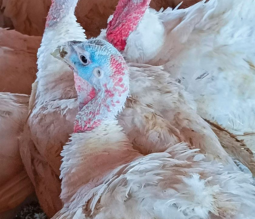
Photo 1. Infraorbital sinusitis.
Photos 2 and 3. Nasal discharge and infraorbital sinusitis.
This is another infection of the upper respiratory tract whose most characteristic symptoms are submandibular edema, tracheal collapse due to distortion of the tracheal rings (softening), and oculo-nasal secretions and sneezing.
Photo 4. Submandibular edema. Stains of brownish exudate are observed
Treatment
Antibiotic treatments administered by water, injection, or spray have been reported to produce minimal clinical improvement. However, the application of tetracyclines, particularly long-acting because of their high bioavailability in tissues and fluids, has been shown to reduce morbidity and duration of the process.
In three clinical cases diagnosed in the field, the animals exhibited prostration, symptoms of the upper respiratory tract with oculonasal discharges, and submandibular edema with brownish ocular exudate in one of them.
This gram-negative bacterium from the Pastereullaceae family and Pasteurella genus can cause acute clinical cases due to its rapid contagion and systemic dissemination, leading to high morbidity and mortality.
At necropsy, hyperemia is observed mainly at the level of the abdominal viscera and associated veins, petechial lesions, and foci of ecchymosis at the level of the heart, gizzard, and intestinal mucosa.
The chronicity of the disease is possible depending on the strain. In these cases, the lesions are more localized, being able to find sternal bursitis, unilateral or bilateral cuprous pleuropneumonia, and joint synovitis.
Pasteurella multocida can be diagnosed from their isolation from visceral swabs, especially lung or bone marrow, from freshly dead or diseased animals slaughtered for sample collection.
Photo 5. Pulmonary hemorrhage.
Treatment with antibiotics from the tetracycline family reduces the morbidity and mortality of the affected batch.
To guarantee the reasonable use of antibiotics, it is essential to carry out antibiograms. These can be useful for future outbreaks of the same flock.
It is recommended to implement vaccine prophylaxis through autovaccines that allow antibiotic therapy to be limite
Photo 6. Lung with caseous fibrinous exudate on the pleural surface.
Animals infected with Ornithobacterium rhinotracheale exhibit coughing, sneezing, sinusitis, dyspnea, prostration, and decreased feed and water intake.
The most frequent necropsy findings are pneumonia and polyserositis.
For the diagnosis of the disease, it is essential to use laboratory techniques, PCR of tracheal and choanal swabs, or bacterial isolation from the heart and liver because the symptoms and lesions described above are non-specific and compatible with other bacterial infections (Pasteurella y E. coli).
Regarding curative or prophylactic treatments, it should be noted that sensitivity to antibiotics by ORT has been documented as inconsistent, with multiple inconclusive studies, depending on the strains and geographical areas. (Wafaa A. Abd El-Ghany, 2021) and that there are currently no commercial vaccines available.
Mycoplasmas are bacteria that lack a cell wall and are classified within the Mollicutes class, Mycoplasmataceae family.
Symptoms
Diagnosis
MG has no treatment, and infected birds are infected for life.
Photos 9, 10, and 11. Sinusitis, unilateral or bilateral swelling, runny nose, and conjunctivitis are observed
Aspergillus is a filamentous fungus of the Trichocomaceae family. It is the most frequently found causal agent in respiratory lesions with a fungal cause.
From the first days, symptoms can be observed with prostrate animals, ruffled feathers, respiratory distress, and sometimes exudative secretions at the periorbital level.
Photo 12. Polyserositis, complicated by E.coli
Necropsy and diagnosis
Although different antifungal treatments have been proposed for avian aspergillosis, there is no effective therapy.
Disinfection of drinking water and litter with antifungals minimizes the clinical course.
Photo 13. Miliary lesions in turkey lung, 4 days
Preventive therapy, such as vaccination plans and the supervision of their correct application, are the treatments to be taken into account to minimize regular antibiotic therapies.
Biosafety and management continue to be understood as fundamental elements of prevention.
It may interest you: Key points in the broiler turkey hatchery process
[/register]
PDF
