Animal Nutrition
IDENTIFICATION OF MYCOTOXIN RELATED LESIONS IN SLAUGHTER HOUSES
To read more content about AviNews-América Latina Sep 2015
Animal Nutrition
To read more content about AviNews-América Latina Sep 2015
Content available at:
IDENTIFICATION OF MYCOTOXIN-RELATED LESIONS IN SLAUGHTERHOUSES
Frequently, poultry producers worldwide are interested in determining if mycotoxins identified in feed or in ingredients thereof are affecting flock productivity.
In case of companies which have decided to add anti-mycotoxin additives, they have doubts as to whether the extra cost which represents their inclusion in the portion is justified.
Being able to quantify the toxin concentration is a tool which may help to make the appropriate decision, but, unfortunately, their detection in raw materials is characterized by inconsistent results, due to their non-uniform distribution and the errors produced when sampling.
Given all these limitations, a practical approach is to assess the presence of mycotoxin-induced lesions in the slaughterhouse. The identification of these lesions, together with the evaluation of the productive parameters, will allow us to determine if they are causing damage and if preventive plans work.
ORAL LESIONS
Mycotoxins responsible for this lesion pertain to the trichothecene group, and include T2 toxin, MAS (monoacetoxyscirpenol) and
DAS (diacetoxyscirpenol).
In the initial phase, damage appears as yellowish patches which later become ulcers localized in palate, tongue, base of mouth, salivary duct outlet and the beak’s lower margin.
After healing, only small ulcers are left remaining on the affected area. In severe cases, necropsy of the tip of the tongue or of all the organ may occur. Oral lesions are more frequently detected after long feeding periods of time, generally in chicks and hens, however, if mycotoxin concentration is high, lesions will appear earlier. Generally speaking, detection in 3-5 week-old fowls is more difficult.
DIFFERENTIAL DIAGNOSIS
The presence of wheat deposits in the corners of mouths, in regions wherein this grain is used, is a condition that should be ruled out in a differential diagnosis. Unfortunately, inspectors working in slaughterhouses have no time to evaluate this type of lesion since the carcasses go quickly through the processing lines and there is not enough time to open the oral cavity.
Evaluating the presence of mycotoxin-induced lesions is a good approach for quantifying their impact
PROVENTRICULITIS/
PROVENTRICULOSIS
Proventriculitis consists of an enlarged proventriculus and proventriculosis is the inflammation of the inner surface. When carrying out an inspection in slaughterhouses, the easiest lesion to identify, without having to open the organ, is proventriculosis. The main mycotoxin responsible for this lesion is cyclopiazonic acid, produced by the fungus Aspergillus flavus, the major aflatoxin producer.
DIFFERENTIAL DIAGNOSIS
Other aetiological agents are the biogenic amines present in bone meals, blood, meat and feathers. The extent of the lesion depends on the amount added to the feed and the type of processing the meals have been subjected to.
Also, pathogens such as Marek virus, Reovirus and Infectious Proventriculitis virus associated with Gumboro virus may cause it.
GIZZARD EROSION
This is a very common lesion which may be caused by T-2 toxin, MAS and/or DAS. Its caustic effect is responsible for the necrotic damage observed in the mouth, gizzard and intestines.
DIFFERENTIAL DIAGNOSIS
Copper sulphate, biogenic amines present in the above-mentioned meals and the use of fish meal may cause a certain degree of organ erosion.
In the past few decades, several scientific publications have reported cases of gizzard erosion produced by Adenovirus A, serotype 1 (FadV-1) in Japan, Poland, Italy and Germany.
In the USA and other countries, these erosions have been reported in 18 day- old chicken embryos and in newborn chicks, apparently associated with hatchery management errors. We frequently visit commercial broiler chicken farms in Latin America and Asia wherein gizzard erosion is reported in the hatchery during hatching and it turns out that two or three weeks later, they almost completely disappear, without affecting production parameters.
Aspergillus flavus produces cyclopiazonic acid which may be responsible for proventriculosis
Intestinal hemorrhages and enteritis may be caused by mycotoxins such as T2 toxin, DAS and/or MAS, characterized in that they produce petechial and ecchymotic hemorrhages that disseminate throughout the intestines.
INTESTINAL HEMORRHAGES/ENTERITIS
DIFFERENTIAL DIAGNOSIS
There are several causative agents, including viral infections (Reovirus), bacterial pathogens (Escherichia coli,
Salmonella) and nutritional factors. In differential diagnosis, it is important to rule out the presence of hemorrhages of death throes, a post-mortem change appearing in some fowls while being slaughtered by neck dislocation, jugular vein cut or carbon dioxide suffocation.
Hemorrhages may appear as petechiae or ecchymosis in the duodenum. These changes are induced by blood accumulation in the lymphoid aggregates situated in the duodenum, which disseminate throughout the intestines and form part of the intestine-associated lymphoid tissue, and includes anatomical structures such as cecal tonsils, Peyer’s patches and Meckel’s diverticulum.
Typically, mycotoxin-induced hemorrhages are distributed throughout the intestines (small and large intestines) and they do not circumscribe to the duodenum such as hemorrhages of death throes.
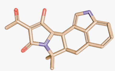
Figure 2. Cyclopiazonic acid structure which may be responsible for proventriculosis
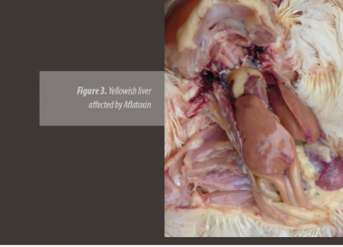
PALE, YELLOWISH AND/OR FATTY LIVERS
Generally, the liver of healthy young fowls, chickens and pullets, is brown colored with a compact aspect.
The “target” organ of aflatoxin is the liver, causing a yellowish discoloration due to lipid accumulation as a result of the harmful effect on the amino acid synthesis ability, which are necessary for fat mobilization in the liver.
DIFFERENTIAL DIAGNOSIS
Layers\\ Most production healthy layers have a pale or yellowish coloration at an hepatic level. However, sometimes, excellent egg-producing hens are detected showing a chestnut brown colored liver.
These differences are important when making a differential diagnosis since physiological changes occurring in a layer may be mixed with the damage caused by mycotoxins.
Chickens\\ In young fowls, yellow or pale livers may be the result of fatty liver and kidney syndrome, a disease occurring from 1 to 4 week-old chicks and which is associated with biotin deficiency. Affected chickens show a growth depression and fatty infiltration in the liver, kidney and heart, without the presence of the characteristic signs of biotin deficiency.
On the other hand, the presence of pale and yellowish livers has been associated with the use of bad quality fats in feed.
When evaluating the color of the liver of chickens in the slaughterhouse, once the liver is mixed with ice, it will look somewhat pale if compared to the liver of fowls examined short after the slaughter. If chickens are completely bleeded after euthanasia in the farm, the liver will be paler than in the case of slaughtered fowls which are not bled.
Fusarium responsible for fumonisin production
FATTY LIVER IN HENS
Fatty and hemorrhagic liver syndrome is characterized by the presence of excessive fat deposits and hemorrhage in the abdominal cavity with enlarged, friable and pale/yellowish livers. This clinical profile is considered one of the most common metabolic disorders reported during the production stage. Under normal conditions, healthy flocks show fowls affected by fatty liver, mostly after 45-50 weeks of age.
When this fatty syndrome is caused by mycotoxins, mostly aflatoxin, the liver has a yellowish aspect with hemorrhagic petechiae, with no excessive fat deposits being observed.
In recent years, the importance of fumonisin has been highlighted in animal production, therefore it is important to consider that even though it has certain effect on the liver, in published scientific papers, even when 100 ppm levels are used, an important change in liver coloration has not been reported.
The main indication of liver damage is sphingolipid metabolism interference, which is determined measuring blood sphingonine and sphingosine (biomarkers) concentrations.
GALLBLADDER WITH PALE CONTENT
The presence of pale bile has been associated with aflatoxin and is the result of the reduction of amylase, lipase and biliary salt production by the liver
BRUISES
It is a relatively common lesion affecting chickens in farms and carcasses in slaughterhouses in many regions. It is considered an important defect because of its negative impact on the presentation of processed chicken in supermarkets.
DESCRIPTION
The presence of hemorrhages in the muscles and underneath the skin (subcutaneous tissue) is observed when they are slaughtered in the farm or in the slaughterhouse and they are the result of surface wounds produced by an impact which does not rupture the skin.
The color of the bruises indicates the time of their occurrence. If they are recent bruises, they are red, blue or black. If they are old bruises, they are green or yellow.
Bruises may occur during chicken transport as a result of the way in which they are trapped and transported to the processing plants.
The main mycotoxin associated with the condition is aflatoxin, characterized in that it increases capillary fragility, interfering with coagulation time and specifically affecting prothrombin effect.

Figure 4. Hemorrhages in the muscles caused by Aflatoxin in commercial chickens
Aspergillus
ochraceus, is one of the fungi responsible for ochratoxin production
In the USA, experiments have shown that concentrations of only 100 ppb may cause bruises in chicken carcass.
It is important to note that recent scientific reports coming from Brazil indicate that fumonisin is associated with a greater incidence of skin hemorrhages in chickens.
DIFFERENTIAL DIAGNOSIS
Besides mycotoxins, stunning carried out by a gentle electric shock in the slaughterhouse may cause an incomplete bleeding.
On the contrary, if a very strong electric shock is used, the presence of hemorrhages due to artery and blood vessel rupture may be increased.
Infectious anaemia virus and Gumboro virus may cause hemorrhages in the muscles and skin.
Under commercial conditions, subcutaneous or muscle hemorrhages are commonly observed in broiler chickens which, in many cases, are related to certain challenge level with Gumboro virus. Various clinical assays in the field have reported that very virulent Gumboro virus are characterized in that they produce more severe hemorrhages in the muscles and skin than classical strains or variants of the virus.
Intoxications may also play an important role. Medicament mistakes with sulfonamides result in a toxic condition known as hemorrhagic syndrome.
KIDNEYS – URATE DEPOSITS AND SWELLING
Ochratoxin is the mycotoxin which more frequently affects this organ in commercial farms. Also, reports on citrinin effect on kidneys are not very common under commercial conditions.
DIFFERENTIAL DIAGNOSIS
Nephropathogenic infectious bronchitis virus strains, excessive dietary calcium levels and lack of water may also cause kidney damage.
For decades, tropism for the kidneys of some Bronchitis strains such as Gray, Holte, Florida 88 has been recognized. Chinese strains such as QX and Q1 have been added to this group and they have already been reported as causative agents of this type of lesions in some areas of China, Europe and Latin America
The following table includes the most important organs which must be weekly or monthly evaluated in slaughterhouses in order to detect the effect of mycotoxins on fowls.

It is important to emphasize that even the most efficient anti-mycotoxin additives existing in the market do not adsorb 100% of these toxins, therefore, certain degree of lesion may always be expected if the level of feed contamination is high.
Samples must be taken regularly of tissues wherein it is macroscopically suspected that mycotoxins are the causative agents in order to fix them in formalin and send them to the histopathologist with the aim of reconfirming the diagnosis.
If practical, during the evaluation of the organs, the inclusion of the bursa of Fabricius which may suffer atrophy as a consequence of mycotoxicosis, will allow for the evaluation of the immune system status. Although the fact that other pathogens may cause small bursas should be taken into account.


RECOMMENDED