Content available at: Español (Spanish)
Over the last three years in the United States, there has been significant cases of Ornithobacterium rhinotracheale (ORT), especially in turkeys, making this disease worrying. If in 2015 it was casuistically put it in number 7 of the most frequent diseases in turkeys, in 2016 it jumped to the fourth position and then to third place by 2017. 
- Stunted growth
- Decreased egg laying
- Mortality in severe cases for all types of birds, especially in turkeys and broilers.
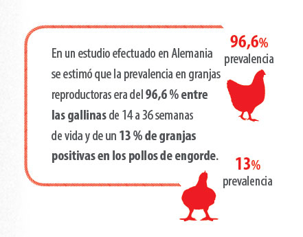
It was first identified in 1994 and the rise in cases due to its highly contagious characteristic is increasing year by year. It is more than likely that the pathogen has been overlooked for years due to lack of identification and that cases of respiratory diseases have not been well diagnosed. Seven serovars have been identified – Empel et al. 1997–, with type A being the most frequent at 95% of identifications.
In turkeys, field cases are usually more frequent between 23 and 42 weeks of age and in broilers between 4 and 6 weeks. The incubation period of the disease stands between 48 and 72 hours.
The airway is the fastest and most common route of transmission, although it can also be via the transovarian route. There are discrepancies regarding its pathogenicity, varying according to the isolates studied and the state of the affected birds. The clinical signs produced, its duration, morbidity and mortality are extremely variable, exacerbated by poor management (inadequate ventilation, high density of animals, poor litter quality, etc.) and by other persisting diseases.
A more severe respiratory condition has been observed when associated with Escherichia coli. The respiratory symptoms observed are:
- Rhinitis
- Coughing up bloody mucus
- Inflammation of the infraorbital sinuses
- Wattle edema
- Tracheitis
- Conjunctivitis or dyspnea
- The nervous signs are due to instabilities, as a results of an affection in the head and the locomotor symptoms to arthritic processes.
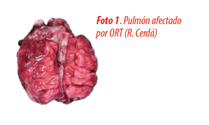
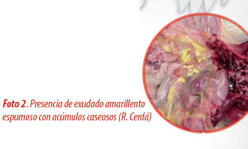
- Lesions seen are limited to the infraorbital sinuses, tracheae, air sacs, lungs, and femoro-tibi-tarsal joints. -Photo 1-.


Diagnosis in the field is usually difficult as it depends on the symptoms and post-mortem lesions. The laboratory diagnosis with positive confirmation from ELISA or isolation of the bacteria fully helps us to confirm the diagnosis. But the culture is complicated due to the microaerophilic and antibiotic conditions present in the culture that are needed so as not to be masked by other bacteria present. Among the control measures for the disease are serological monitoring, which is highly recommended to carry out the real-time PCR technique, isolation, and the respective antibiogram. The ELISA technique is recommended as very suitable for the diagnosis of infection in birds.
Administration of doxycycline in drinking water and chlorotetracycline in compound feed reduces the action of the bacteria. Administration of amoxicillin (250 ppm) for 4 to 7 days also offers good results in reducing mortality from the bacteria. The use of macrolides such as tylosin and tylvalosin are another good way to act against the bacteria. There is inactivated vaccine on the market and the possibility of making auto-vaccines to help control the disease.
More research is needed to better understand this disease and the options to control it, since its interrelation with other viral agents present in poultry farms (especially avian bronchitis and Mycoplasma gallisepticum) may favor its pathogenicity.


