Content available at: Indonesia (Indonesian) Melayu (Malay) ไทย (Thai) Tiếng Việt (Vietnamese) Philipino
Avian Metapneumovirus (aMPV)
Respiratory diseases are a constant challenge for poultry producers and veterinarians since they do not show pathognomic signs, making diagnosis more complex.
- The Avian Metapneumovirus (aMPV) is an important pathogen in respiratory diseases but is frequently neglected.
- It can affect the respiratory and reproductive systems, facilitating the development of other diseases like colibacillosis, which is the most common co-infection in broilers, while in turkeys, it is Ornithobacterium rhinotracheale.
- Mycoplasma gallisepticum infection can prolong the viral replication, but as a secondary infection, aMPV can delay M. gallisepticum infection.
Distribution and epidemiology of aMPV
The aMPV affects turkeys and chickens and can also be found in guinea fowl, ducks, and pheasants.
It is an enveloped negative-sense RNA virus included in the genus Metapneumovirus of the Pneumoviridae family.
The global impact of aMPV is significant. Six viral subtypes of aMPV are recognized worldwide, each with its unique distribution.
- Subtypes A and B are found in Europe, Brazil, and the African continent, while subtype C has been identified in the United States, Canada, China, France, and South Korea.
- The D subtype was only reported in France, and the two new subtypes are found in the United States and Canada in wild birds (black back gull and Monk parakeet chicks).
- The genetic differences among subtypes are based mainly on the glycoprotein (G) variability that also affects replication in the target cell and, consequently, the pathogenicity in the host.
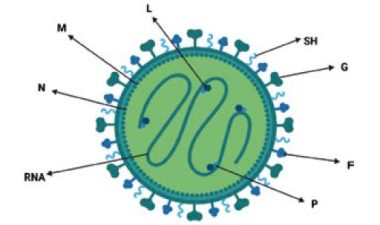
Schematic figure representing aMPV: (G) Glycoprotein, (F) Fusion protein, (SH) Small hydrophobic protein and other structural proteins, (M) Matrix protein, (N) Nucleocapsid protein, (P) Phosphoprotein, (L) RNA-dependent RNA polymerase and RNA strand.
Currently, subtype B is the most prevalent worldwide. However, subtypes A and B are causing outbreaks in several states in the USA, following a prolonged period without aMPV detection.
The recent introduction of aMPV-A and -B into the US has coincided with the increased spread of H5N1 HPAI by migratory waterfowl.
- Whether these two occurrences are causal relationships or random effects remains to be determined.
- The shift in the predominance of aMPV subtypes from A to B and the increased detection of aMPV-A in North America in recent years are significant and may reflect ongoing evolutionary dynamics and changing
epidemiological patterns.
The presence of aMPV has been confirmed in wild migratory birds in several countries, indicating that this factor must be considered in epidemiological studies, as well as seasonality and biosecurity to prevent it. Horizontal transmission through aerosol is the most common route of infection, and no vertical transmission has been reported.
Pathogenesis
- Metapneumovirus has a high tropism for the upper respiratory tract, infecting sinuses, larynx, and trachea.
- It causes cessation of ciliary movements or ciliostasis and even complete loss of these cilia (desciliation).
- These lesions cause mucus not to be removed and accumulate in the passages and cavities, causing the main sign of this disease, the swollen head.
- It causes severe respiratory infection in turkeys, better known as Turkey Rhinotracheitis (TRT).
- In laying hens can cause a drop in egg production, and it has been isolated from cockerels’s testes, causing reduced fertility together with the Infectious Bronchitis Virus.
Common symptoms include:
- Sneezing.
- Nasal and eye discharge.
- Conjunctivitis.
- Submandibular edema.
- Infraorbital sinus swelling.
- Cracking.
- Rales.
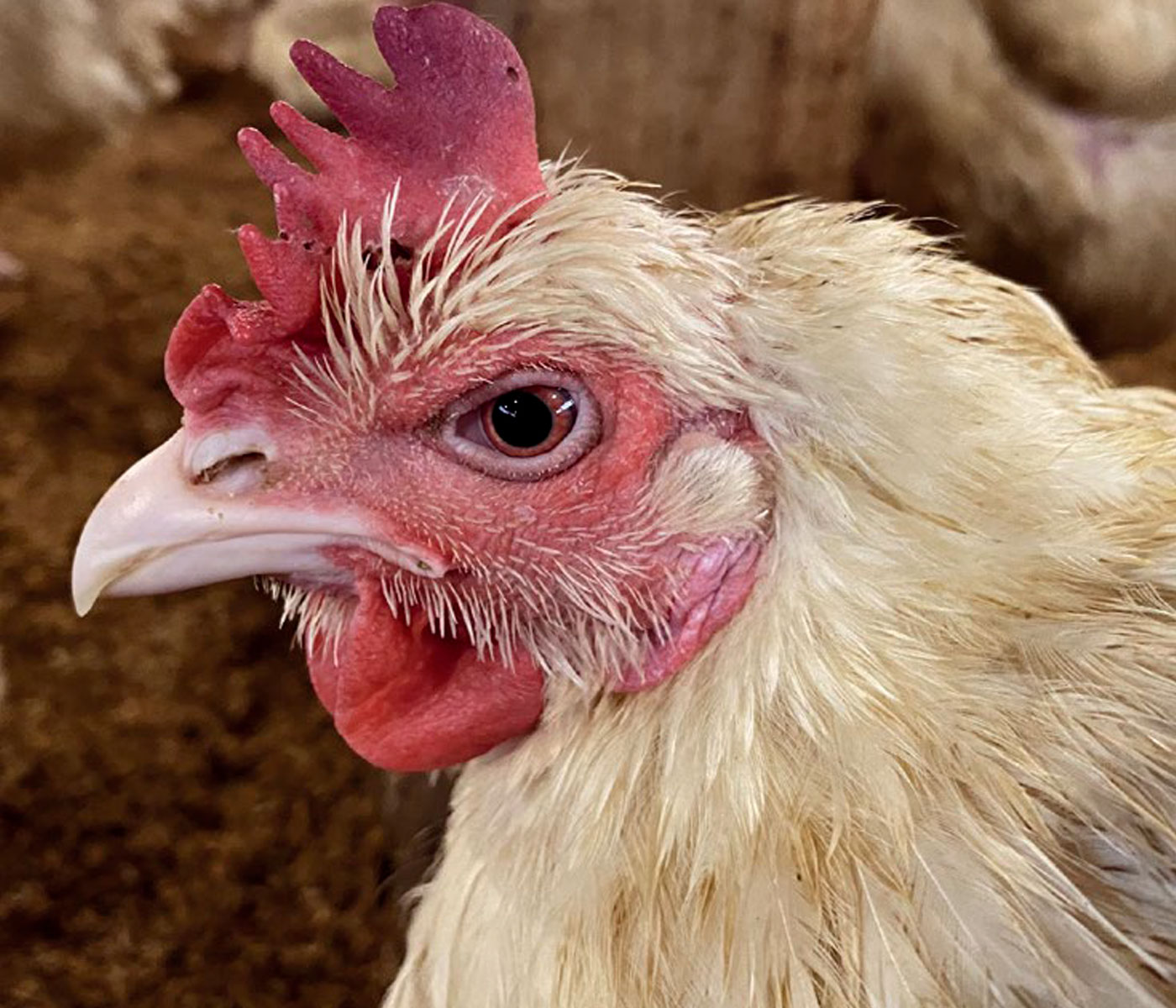
In chickens, it causes swelling of the periorbital and infraorbital sinuses, torticollis, disorientation, and opisthotonos. The clinical manifestation may progress to reddening of the conjunctiva with edema of the lacrimal gland.
- Between 12 and 24 hours, the birds show a subcutaneous swelling on the head, which starts around the eyes, increases under the entire head, and then descends to the submandibular tissue and back of the neck.
- After three days, chickens may show neurological signs such as apathy and torticollis.
- The permanence period of aMPV is only 4 to 7 days, which impairs virus detection for molecular diagnosis.
Vaccination and selective pressure
- Vaccines from subtypes A and B are available, and both products provide good cross-protection.
- Currently, no licensed vaccine is available for use in the USA.
- A possible selection by vaccine pressure has been reported in the past two decades.
- The heterologous protection capacity between vaccines of both subtypes is demonstrated, although it is susceptible to viral escapes.
- Then, vaccination must always be paired withbiosecurity and virus surveillance for strategic control.
There are contradictory reports regarding the evolution of aMPV.
Some studies report that aMPV is a relatively slow-evolving virus compared to other avian RNA viruses, and others estimate that its rate of viral evolution is within the normal range.
- Nonetheless, viral evolution is based on both the pressure exerted by vaccine programs and the type of host and environment; consequently, multiple strains of the same subtype can phenotypically circulate in distinct regions of the globe.
- The genetic diversity in the study sequences highlights the urgent need to carry out more whole genome sequencing of this virus to better understand the variants circulating in the field and the evolution of this virus over time.
The phylogenetic relationships among the aMPV-B strains, reconstructed using the maximum likelihood method in the Molecular evolutionary genetics analysis (MEGA X) software, showed that aMPV-B has evolved in Europe since its first appearance.
- The 40% of aMPV-B viruses analyzed were identified as vaccine-derived strains, sharing phylogenetic similarities and displaying high nucleotide similarity with live commercial vaccine strains licensed in Europe.
- The remaining 60% were considered field strains because they clustered separately and showed a low nucleotide identity with vaccines and vaccine-derived strains. Contrary to vaccine-derived strains, field strains tended to cluster based on their geographical origin and irrespective of the host species where the viruses had been detected.
Diagnostic tests and discovery of new viral strains
All experts recommend monitoring birds by serology to detect antibodies by ELISA and detect the virus to identify the prevalence of the subtype and determine the best vaccine to use.
The following commercial ELISA kits are available:
- Idexx Avian Pneumovirus antibody test kit to detect subtypes A, B, and C
- BioChek Avian rhinotracheitis (ART) antibody test kit to detect subtypes A and V
AMPV subtypes A, B, C, and D can be detected by traditional real-time PCR or RT-qPCR. However, virus isolation and genome sequencing of the G gene region can be necessary to identify subtypes.
- The molecular characterization of aMPV and the differentiation between vaccines and field strains through G gene sequence analysis can be valuable tools for correct diagnosis.
- They should be routinely applied to better address the control strategies.
Common samples for analysis are oropharyngeal, nares, and sinus cavity swabs that should be submitted in transport media. For asymptomatic birds, the nares are the most reliable sample site for molecular diagnosis of aMPV, with the choanal cleft and trachea being secondary options. Symptomatic birds normally have detectable virus in the choanal cleft, nares, and trachea.
- Maintaining a cold chain from the point of collection through transport to the diagnostic lab is critical.
- If shipping may take more than 24 hours, samples should be frozen at—80 oC and shipped express on dry ice.
The control of aMPV requires correct identification of the agent, epidemiological surveillance, effective diagnosis, adequate immunoprophylactic programs, and constant biosecurity.
A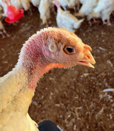
B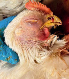
Figure 1. (A) Turkey showing swollen infraorbital sinuses, ocular discharge, and conjunctivitis (courtesy of Dr. Ashley Mason); (B) five-week-old chicken showing swollen head, swollen infraorbital sinuses, plugged nostrils, and opaque crusted ocular discharge (courtesy of Dr. William McRee). Source: Luqman et al., 2024. Viruses 16 (4): 508.

