Content available at: Indonesia (Indonesian) Melayu (Malay) ไทย (Thai) Tiếng Việt (Vietnamese) Philipino
Modern poultry lines are characterised by high performance and specialisation. Advances in genetics, feeding, management, diagnosis, environment and breeding have significantly improved production parameters.
While today’s birds are more efficient and productive, they are also less hardy than in the past, making them more susceptible to disease.
- Because of this increased susceptibility, modern production requires strict biosecurity measures and efficient vaccination schedules.
- More and better vaccines are now available, however, the immune system of the birds needs to be in optimal working condition.
Due to the multifactorial aetiology for immunosuppression, diagnosis is not always straightforward, requiring a clinical history, necropsies and complementary laboratory tests if necessary.
- It is very important for the field clinician to be aware of the macroscopic changes in the immune system that can be observed during necropsy. This guides the diagnosis and facilitates sampling.
In the following, the most relevant aspects of immunosuppression-oriented pathology of the immune system are discussed.

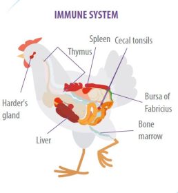
The avian immune system consists of primary and secondary lymphoid organs.
- During the embryonic stage, undifferentiated cells migrate from the yolk sac to the bone marrow, thymus and bursa of Fabricius.
- In these organs the cells differentiate into T- or B-lymphocytes where they express surface markers and by negative selection those lymphocytes that are not useful are eliminated.
- They then migrate to secondary lymphoid organs such as the spleen, caecal tonsils, Harder’s gland, mucosa-associated lymphoid tissue, and germinal centres in connective tissue.
The interpretation of lesions in lymphoid organs requires taking into account the age of the birds and vaccination schedule, as primary lymphoid organs atrophy when birds reach sexual maturity and most routine vaccines cause changes in the lymphoid organs.

It is present only in birds, located in the dorsal part of the cloaca, connected to the intestine by a duct.
- The bursa of Fabricius is the site of differentiation of B lymphocytes and captures antigens when the bird defecates, as the smooth muscle layer of the intestine continues into the bursa of Fabricius so that the contraction of the muscle makes the bursa function as a suction knob.
Inside the bursa of Fabricius there are major and minor folia, these folia are lined by a columnar epithelium and contain the lymphoid follicles supported by a matrix of connective tissue.
- Each lymphoid follicle is made up of a cortex and medulla, separated by a layer of cortico-medullary cells that continue with the cells of the epithelium lining the folia.
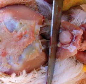
The bursa of Fabricius, in the absence of infectious agents or immunosuppressants, should be present until 12 to 14 weeks of age, at which time it begins to involute, so that by 20 weeks only traces remain.
- In production birds, the use of vaccines, mainly against infection of the bursa of Fabricius, causes atrophy before this time.
- The involuted bursa of Fabricius appears macroscopically as a small, hard, yellowish-white nodule.
- Microscopically, the folia are small, the follicles have lost the differentiation between cortex and medulla, the epithelium shows folds and cysts.
In 1-day-old chicks, clusters of heterophils are frequently found in the subepithelial tissue; these are foci of extramedullary granulopoiesis and are common in various tissues of the chick.
THYMUS
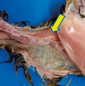
The thymus in birds is located along the neck and is composed of 6 to 7 lobes running parallel to the jugular veins and the vagus nerve.
- T-lymphocytes differentiate in the thymus.
- Macroscopically, the lobules of the thymus are seen with small lobules and on sectioning, a cortex and medulla can be distinguished.
In the absence of infectious or immunosuppressive agents, the thymus should remain until 15 to 17 weeks, after which time it begins to involute so that by 30 weeks only vestiges remain.
- Histological examination of involuted thymus shows loss of the cortex and fibrosis of the medulla.
BONE MARROW
Only non-pneumatic bones such as the femur and tibiotarsus have bone marrow, it is considered a primary and secondary lymphoid organ as bone marrow is the source of:
- Undifferentiated cells that migrate to the thymus and bursa of Fabricius in embryonic life and;
- On the other hand, they are a source of these same cells in adult animals or in the repopulation of the thymus and bursa following severe damage with loss of lymphocytes.
SPLEEN
The spleen is attached to the gizzard and proventriculus by its visceral side. It is a secondary lymphoid organ, made up of a capsule of connective tissue and trabeculae on which it is supported:
- The germinal centres (white pulp) and arterioles.
- The dendritic cells.
- Red blood cells (red pulp).
The spleen in young chicks is a centre of granulopoiesis and in older birds a centre of antigen presentation.
LYMPHOID ORGAN ATROPHY
In addition to the age-related involution of birds, there are many factors that cause lymphoid atrophy.
Birds are subjected during the production process to stress factors such as:
- Heat.
- Cold.
- Handling.
- Vaccinations.
- Feed restrictions.
- Selection and regrouping among others.
Which trigger the secretion of glucocorticoids which cause apoptosis in lymphoid cells.
APOPTOSIS PROCESS
The process of apoptosis is normal in birds during negative selection processes of clones that are not useful. A histological slice of lymphoid organs shows apoptosis, which is described in texts as ‘starry sky’.
- This is considered physiological unless the apoptosis is such that the size of the organ is decreased.
On the other hand, mycotoxins are considered to be immunosuppressive agents that cause atrophy of lymphoid organs, the mechanism of atrophy following two pathways:
- They inhibit protein synthesis and nucleic acid replication, which are necessary for lymphoid cell differentiation and proliferation and;
- Birds reject feed, which generates stress and glucocorticoid release.
If decreased lymphoid organ size is observed, mycotoxins should be considered in the differential diagnosis.
In the case of the thymus, atrophy due to abundant apoptosis occurs in cases of infection with infectious anaemia virus. This vertically transmitted virus targets T lymphocytes and bone marrow germ cells.
Birds under 5 weeks of age show severe atrophy of the thymus cortex, decreasing considerably the size of the thymus, sometimes at necropsy the thymus may go unnoticed.
NECROSIS AND INFLAMMATION
In addition to apoptosis, necrosis also leads to atrophy of lymphoid organs, but in this case the inflammatory process may transiently increase the size of the organ.
- In the case of the bursa of Fabricius, the best known agent is the bursa of Fabricius infection virus (IBF), also known as Gumboro disease. The changes that the virus causes in the bursa of Fabricius have been well characterised.
- Lymphoid cell necrosis with oedema between follicles and germinal epithelium, as well as heterophilic infiltration are the first changes.
- When infection is caused by very virulent strains (hot strains) haemorrhages occur.
- During the first 3 to 5 days, the bursa of Fabricius increases in size and after a week there is proliferation of cortico-medullary cells and fibrosis with a decrease in the size of the bursa, the epithelium is folded and cysts can be seen.
These changes are not exclusive to IBF, as other agents such as Marek’s disease virus (MDV), Reovirus and Newcastle disease virus (NDV) can also cause necrosis and inflammation.
- For this reason, the diagnosis of IBF by histopathology must be accompanied by antibody determination by ELISA or virus serum neutralisation.
A particular case is IBF virus infection by variant strains, as they do not cause necrosis and inflammation and the bursa of Fabricius only atrophies, these viruses cause lymphoid cell death by apoptosis.
- In the absence of secondary infections, the bursa of Fabricius may recover after 8 weeks, in which case areas of regeneration characterised by intense basophilic staining can be seen in the vicinity of depopulated follicles.
Regeneration depends on the absence of secondary infections, if there is a bacterial complication or cryptosporidium infestation the bursa of Fabricius has a fibrinous to fibrinocaseous exudate that may fill the bursa.
LYMPHOPROLIFERATIVE DISEASES
In birds, Marek’s disease (MD) and Lymphoid Leukosis (LL) viruses cause lymphoid cell neoplasms (lymphomas) occurring in various organs.
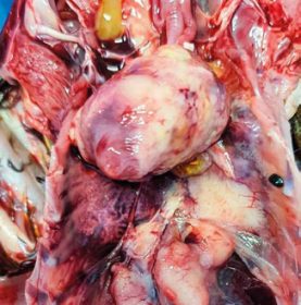
- Necrosis and inflammation of the thymus is rare, usually associated with the application of emulsified vaccines, in which case a granulomatous reaction is observed.
- On the other hand, necrosis of lymphoid cells and hyalinisation of arterioles can be observed in cases of NDV in its velogenic presentation or in Avian Influenza (AI), particularly with the H5N2 virus.
- Most viral agents that cause immunosuppression with lymphoid cell necrosis or apoptosis result in spleen cell depletion, and when the immune system is constantly stimulated by other viruses and bacteria, lymphoid hyperplasia occurs.
- With the naked eye, the cut surface shows white stippling, which microscopically corresponds to nodules of lymphoid hyperplasia. In cases of NDV or AI, areas of infarction with hyalinisation of arterioles are present.
Marek’s disease
Marek’s disease has 5 presentations depending on the site of T-cell infiltration. With the exception of the cutaneous presentation, all other presentations are nonproductive transforming, that is, complete virions are not produced and the infected cell becomes neoplastic.
Clinical manifestations depend on the organ infiltrated, thus birds present:
- Blindness at ocular presentation.
- Paralysis or pendulous crop in the nervous presentation.
- Various disorders including diarrhoea, dyspnoea, renal failure, ascites, etc.
The above depends on the organ infiltrate in the visceral presentation. In this case the organs present firm, white nodules in most cases. Muscular presentation is the rarest.
Lymphoid Leukosis
Within the differential diagnosis with LL, it should be considered that LL is only visceral, so it is important:
- The examination of nerves and bursa of Fabricius.
- The appearance of the neoplastic cells.
- The age of the birds.
Lymphoid leukosis presents intrafollicular infiltration (within the lymphoid follicles) in the bursa of Fabricius and the tumour is frequent. While in cases of MD the infiltration is interfollicular (outside the follicle, occupying the connective tissue between the epithelium and follicles) and on the other hand the presence of nodules is less frequent.
In recent years, molecular tests have provided valuable data in the diagnosis and epidemiology of immunosuppressive viral diseases, however, it should not be lost sight of the fact that a positive PCR test result does not always have diagnostic value.
For example: in the case of common infectious anaemia virus in six to seven week old broilers or 10 week old replacement pullets. At this age, virus infection does not cause immunosuppression, so the presence of virus genetic material in the sample alone is not necessarily the origin of the observed picture and other possibilities must be ruled out.
FINAL CONSIDERATIONS
- The interpretation of lesions in the lymphoid system, as well as the repercussions in the flock, requires a detailed clinical history, a correct selection of the birds and complementary serological tests are very helpful.
- Waste or selection chickens are not always the most representative of the flock. Since in these birds the lymphoid organs generally show atrophy because their feed consumption is always lower.
- Also, since lesions in the lymphoid system can be caused by several agents, it is necessary for the clinician to be familiar with serological tests and the results obtained.
