Content available at: Español (Spanish)
For some years now, typhlohepatic parasitic infestation or simply Histomoniasis, has been observed not only in turkey flocks, but also uncommonly in broiler breeders in Europe. The complication lies in the fact that the effective medicine has been banned in the EU since 2003.
The life cycle of the parasite is linked to the nematode Heterakis gallinarum, which is also a parasite of the ceca of birds.
Their eggs are highly resistant to environmental conditions and release the protozoan into the cecum where it multiplies by simple bipartition.

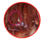
Photo 1. Liver lesion Histomoniasis
As a next step, Histomona meleagridis invades the cecal wall and reaches the liver through the bloodstream.
Another theory being considered is the transmission by a more direct route, i.e: cloacal contact. This may explain the rapid transfer of protozoa from one bird to another during the clinical course of the disease.
The incubation period of the parasite is 7 to 10 days and one of the first characteristic clinical signs is a sulfuric yellow diarrhea, as a consequence of the inflammatory process of the cecum.
Later, anorexia, drowsiness, ataxia, and morbidity start to appear in the birds.
The evolution in birds is usually poor, with a very slow increase in mortality of affected birds two weeks after onset of symptoms.
Cecal lesions affect one or both ceca, and may damage either part or the entirety of the ceca.
The cecal walls also thicken and congest.
In most cases, cecum size is increased as a consequence of the abundant exudate secreted by the mucosa.
At the opening of the cecum, ulcerative, necrotic-caseous lesions and a thick yellowish layer are visible. This can lead to perforation of the cecum wall and induce peritonitis. In chronic cases, adhesions are seen between the cecum and the proximal intestinal loops.

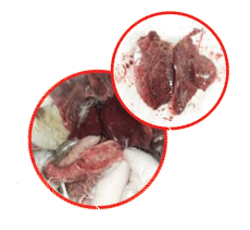
Photo 2. Liver lesion Histomoniasis
Liver lesions are more variable and in some birds they are not observed or are blurred, and may be related to the intensity of the disease process.
Generally, there are pin-shaped necrotic foci with growing edges and a depressed center. Their number in the liver is variable and their size can range from a few millimeters to several centimeters in diameter, giving the liver a mottled appearance. Hepatic hypertrophy and discoloration are also sometimes seen.
The diagnosis based on the observation of the lesions in the two organs is pathognomonic. The presence of the parasite must always be confirmed by microscopic examination, especially in the ceca.
In the EU, effective molecules are banned in the fight against histomoniasis in poultry. Today, paromomycin sulfate is being used for treatment through the use of an exceptional prescription for therapeutic vacuum.
This active ingredient is an oligosaccharide antibiotic from the group of aminoglycosides indicated in human and veterinary medicine for the treatment of intestinal infections caused by amoebae and cryptosporidia.
The prophylaxis of the disease is based on biosecurity and land that has been occupied by chicken and turkeys should never be reused, even at different times.
It is also important to avoid contamination of feed or water with feces to avoid transmission between birds. Deworming must also be considered to combat Heterakis gallinarum and break the biological cycle.
PDF


