Content available at: Español (Spanish)
Much research has been done in recent years on the muscle tissues of poultry, especially due to recent problems of meat quality in the poultry sector. All of this has been particularly related to the rapid development of broilers, determined by advances in genetics and nutrition.
However, these muscular changes, which could be classified as myodystrophies (which occur with the intracellular hyalinosis of the actin and myosin myofibrils), seem to affect the muscles in different ways, which is why we ask ourselves:
To answer this question we have reviewed the histology and general pathology of fiber. Coming to some interesting conclusions, or at least curious. For this purpose, we have divided our work into three parts:
- Firstly, we are going to see the peculiarities of the histology of the skeletal muscle of birds.
- A second part with a general study of myodystrophies in animals.
- Finally we focus on the study of meat problems and their relationship with avian muscles.
Muscular degenerations or myodystrophies comprise a series of alterations of very diverse etiology, but all of them are characterized by a common lesion; INTRACELLULAR HYALINOSIS, that is, the appearance of proteinaceous, intracellular deposits that can be homogeneous, translucent, eosinophilic, structureless or somewhat refractive in nature. Its name comes from the term “hyalos” or glass for its appearance (T. Kitt & LC Schulz: Lehrbuch der Allgemeinen Pathologie für Tierärzte und Studierende der Tiermedizin. Ferdinand Enke Verlag, Stuttgart, 1982)
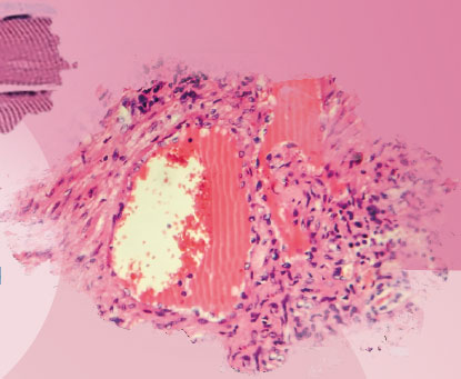
Macroscopically, the muscles appear whitish and dry (waxy): Zenker’s degeneration; Hyalinosis is observed, especially in the adductor muscles of the extremities. Microscopically, hyalinosis is seen first, which later undergoes a Bowman’s discoid fragmentation. Later; and if the cell membrane or sarcolemma breaks, the lesion would be irreversible and there would be necrosis, inflammation, calcification, fibrosis – scarring, and even lipomatous pseudohypertrophy.
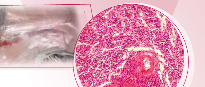
At present the myodystrophies described in birds are classified into six types, according to their causes, which are collected in the classic book by Riddell, C .: Avian Histopathology, AAAP, Pennsylvania, 1987:
- Hereditary
- Dietary or nutritional or white muscle
- Exercise and catch (wild)
- Ischemic: lack of blood supply.
- Toxic: ionophores in turkey (toxic plants)
- Traumatic
TO CONTINUE READING REGISTER IT IS COMPLETELY FREE Access to articles in PDF
Keep up to date with our newsletters
Receive the magazine for free in digital version REGISTRATION ACCESS
YOUR ACCOUNT LOGIN Lost your password?
