Emerging technologies for identifying woody breast syndrome
Contenido disponible en: Español (Spanish) Português (Portuguese (Brazil))Although primary breeders are already actively working to decrease the frequency of myopathies […]
Although primary breeders are already actively working to decrease the frequency of myopathies in general, new research is being generated to automatically detect chicken carcasses affected by myopathies such as "woody breast."
Throughout this brief article we summarize some of the new technologies focused on the identification in vivo or in the process plant of myopathies in an automated way.
Normally, woody breast problems are identified and the carcasses classified manually by carcass palpation by trained employees at processing plants.
Different technologies are currently being investigated to quickly identify this and other myopathies in an automated way:
- Ultrasound imaging
- Photographic imaging to determine the morphology of the breast
- Bio-electrical impedance analysis of the pectoral muscles
- Spectroscopy
- 3D imaging (3D)
- Vis-NIR hyperspectral imaging
Ultrasound
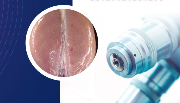
Ultrasound imaging to predict woody breast issues is being investigated using live birds before they are processed. Ultrasound images are correlated with the presence and severity of woody breast problems in already deboned breasts, thus allowing the prediction of their presence.
Photographic Imaging
The generation of photographic images, which helps to accurately determine the shape of the breast in full carcasses, has also been used to correlate the presence and severity of woody breast syndrome. This technology allows the suspect carcasses to be quickly and automatically separated from the normal ones before moving to the deboning station and further processing.
Bio-electrical impedance
Technologies are also being developed for the identification of woody breast immediately after deboning, allowing easy separation of affected breast fillets. Bio-electrical impedance analysis technology works by measuring the ease of electrical signals that get transmitted through the muscle tissue.
Spectroscopy
Spectroscopy is used to measure the composition of chicken meat by detecting the passage of light through the muscle tissue.
Third dimension
Third-dimensional (3D) imaging is based on factors specific to the morphology of breast fillets, which include thickness, volume, area, weight, and density, in order to quickly detect affected breasts.
VIS-NIR
"Vis-NIR hyperspectroscopy" allows the generation of images through the analysis of minute differences in wavelength to be able to separate and discard the breast fillets affected by this myopathy and easily distinguish them from normal breast fillets.


